
Anatomie

Anatomie
E. Genevelle 2001
 Avec cet article, vous avez désormais entre vos mains les outils pour comprendre le jargon des Allgayer, Tawil et Burnel. C’est peu dire et ça va en dégoûter plus d’un. PS: Un grand merci à Pascal Romans (Laboratoire d’Ichtyo-écologie Tropicale et Méditerranéenne. Ecole Pratique des Hautes Etudes. ESA CNRS 8046. Université de Perpignan. 66860 Perpignan cedex) pour l’obtention de la documentation et à Robert Allgayer pour les superbes clichés à balayage.
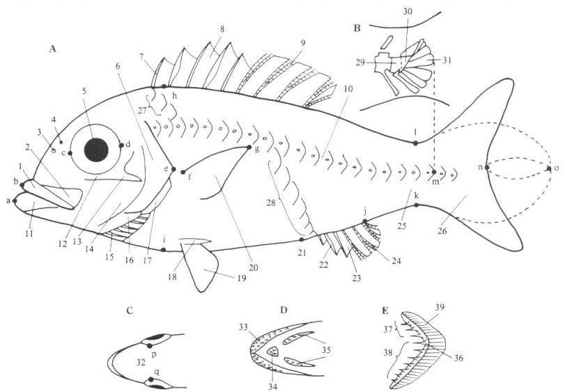
Fig I
|
Position de la bouche. |
 Positions des pelviennes. |
 Types de caudales. |
| 1 | Premaxilla | 6 | Operculum | 11 | Lower jaw | 16 | interoperculum | 21 | Anus | 26 | Caudal fin | 31 | Hypural(s) |
| 2 | Maxilla | 7 | Dorsal fin spine(s) | 12 | Infraorbital bone(s) | 17 | Suboperculum | 22 | Anal fin spine(s) | 27 | Scales above lateral line | 32 | Interorbital space |
| 3 | Anterior nostril | 8 | Dorsal fin | 13 | Infraorbital shelf | 18 | Axillary scale | 23 | Anal fin | 28 | Scales below lateral line | 33 | Upper jaw (teeth) |
| 4 | Posterior nostril | 9 | Dorsal fin ray(s) | 14 | Preoperculum | 19 | Pelvic fin | 24 | Anal fin ray(s) | 29 | Last caudal vertebra | 34 | Prevomer (teeth) |
| 5 | Eye | 10 | Lateral line scale(s) | 15 | Branchiostegal ray(s) | 20 | Pectoral fin | 25 | Caudal peduncle | 30 | First ural vertebra | 35 | Palatine (teeth) |
| 36 | Gill raker | 37 | Gill rakers on upper limb | 38 | Gill rakers on lower limb | 39 | Gill filament(s) |
| Total length: | The greatest dimension between the anteriormost projecting part of the head and the farthest tip of the caudal fin (the caudal rays are pressed together). | Fig. I, a-o |
| Standard length (= body length): | The distance from the anteriormost tip of the snout or upper lip to the posteriormost point of the fold formed when the caudal peduncle is bent | Fig. I, b-m |
| Fork length: | In fishes with a forked caudal fin, the distance from the anteriormost tip of the snout or upper lip to the anteriormost point of the caudal posterior margin | Fig. I, b-n |
| Body: | The greatest dimension, exclusive of fins, and fleshly or scaly structures, which pertain to the fin base. | Fig. I, h-i |
| Preanal length: | The distance from the anteriormost tip of the snout to the center of the anus | |
| Head length: | The distance from the anteriormost point of the farthest margin of the pectoral fin | Fig. I, b-e |
| Caudal peduncle length: | The distance from the base of the last anal ray to the posterior en of the hypural bone | Fig. I, j-m |
| Caudal peduncle depth: | The least depth of the caudal peduncle | Fig. I, k-l |
| Snout length: | The distance from the most anterior point of the snout or upper lip to the front margin of the orbit | Fig. I, b-c |
| Eye diameter or orbital length: | The greatest distance between the free orbital rims | Fig. I, c-d |
| Interorbital width: | The least width between the eyes | Fig. I, p-q |
| Length of the upper jaw: | The distance from the most anterior point of the premaxilla to the posterior end of the maxilla | |
| Mouth width: | The greatest transverse distance across the opening the mouth | |
| Pectoral fin length: | The distance from the extreme base of the uppermost ray to the farthest tip of the fin | Fig. I, f-g |
| Counting scales | ||
| Lateral line scale count: | The number of scales on the lateral line from the scales on the posterior end of the hypural bone | Fig. I, 10 |
| Scales above lateral line: | The number of scale rows above the lateral line from the origin of the dorsal fin, including the small scales, and counting downward and backward following the natural scale row to, but not including the lateral-line scale. | Fig. I, 27 |
| Scales below lateral line: | The number of scale rows below the lateral line is counted similary to that for rows above lateral line. The count is made upward and forward from the origin of the anal fin. In this count, as in the one above the line, the small scales are included | Fig. I, 28 |
| Counting vertebrae | ||
| Vertebrae: | In teleosts the number of vertebrae is counted from the foremost precaudal vertebra articulating with the cranium, to the caudal vertebra attaching to the first ural vertebra. | |
| Counting gill rakers | ||
| Gill rakers: | The number of gill rakers on the outside of the first gill arch, shown by 2 figures separated by a plus sign (+). | Fig. I, E |
| Counting fin rays | ||
| Fin rays: | There are 2 kinds of fin rays, i.e., the spines and the soft rays. Spines are unpaired structures, without segmentation, and designated by large Roman numerals (I; II, …). Soft rays, designated by the Arabic numerals (1, 2, …), are usually, though not always, branched and flexible, and are bilaterally paired and segmented. In a fin containing both spines and soft rays, the count for the spines is separated (e.g., XI-18). If the two sections are joined, a comma is used (e.g., XI,18). | |
| Counting branchiostegal rays | ||
| Branchiostegal rays: | The total number of the rib-like slender bones located below the opercular bones and supported by the membrane | Fig. I, 15 |
| Chez un Tropheus, cela donne ceci:
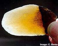
Et les dents ! |
Continuons par une dissection:
|
Radiographies de cichlidés:
 Amphilophus |
 Tropheus |


L’oeil:

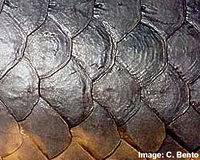
Poursuivons avec les différents types d’écailles que l’on trouve chez les poissons
 Cosmoïde |
 Cosmoïde |
 Ctenoïde |
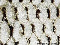 Cycloïde |
|
|
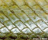 Ganoïde |
 Placoïde |
|
Sources:
Masuda H., Amaoka K., Araga C., Uyeno T. and Yoshino T., 1984. The fishes of the Japanese Archipelago. Vol 1. 431pp. Tokay University Press.
Datz Sonderheft – Tangajikasee
BOULENGER G.A. – Catalogue of the freshwater fishes of Africa in the British Museum – Vol III / 1915
NELISSEN M. H. J – A taxonomic revision of the genera Simochromis, Pseudosimochromis and Tropheus (pisces, cichlidae) – Musée Royal de l’Afrique centrale. Tervuren, Belgique – Annales – série N°8. Sciences Zoologiques N° 229 – 1979
POLL M. – Révision de la faune ichthyologique du lac Tanganyika – Annales du Musée du Congo Belge – C. Zoologie. Poissons, Reptiles, Amphibies – Série I, Tome IV Fascicule 3. – P 141-364 / 1946


